RADIOLOGY
Radiology is an examination to diagnose and see certain medical conditions experienced by patients, using various machines and examination techniques such as X-rays, magnetic fields, sound waves, and radioactive liquids. The main purpose of this radiological examination is to help diagnose and treat disease by providing timely and accurate information from radiological test results.
At Virtu DigiLab, you can get a complete radiology examination service with fast and accurate examination results. Supported by sophisticated and high-quality medical equipment, Virtu DigiLab is able to provide the right diagnosis results.
RADIOLOGY SERVICES AVAILABLE
UNDERSTANDING AND PREPARING YOUR PROCEDURES
Do not have to. X-ray examination is required according to the doctor’s advice and requests.
X-ray examination has many benefits for body health examinations, such as:
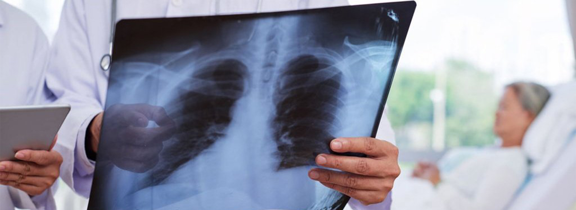
- Lung problems, such as tumors/cancer, infections, collection of air in the space around the lungs (pneumothorax), and other chronic lung conditions, such as emphysema or cystic fibrosis.
- Pulmonary-related heart problems. A chest X-ray can show changes or problems in the lungs that the problem originates from the heart. For example, fluid in the lungs (pulmonary edema) is the result of congestive heart failure.
- Heart size and shape. Changes in the size and shape of the heart can indicate heart failure, fluid around the heart (pericardial effusion), or heart valve problems.
- Blood vessel. The location of the great vessels close to the heart, aorta, pulmonary arteries, and veins is seen on X-rays. That’s why conditions like aortic aneurysm, or other blood vessel problems as well as congenital heart disease can be seen.
- Calcium deposits. A chest X-ray can detect the presence of calcium in the heart or blood vessels. This indicates damage to the heart cavity, coronary arteries, heart muscle, or the protective sac that surrounds the heart.
- Broken ribs or spine.
- To determine the presence / presence of foreign bodies in the lungs, or chest cavity.
The process of taking a chest X-ray does not take more than 10 minutes.
Examination of the chest chest X-ray preparations that need to be done only wear loose clothing and you are asked to inhale as hard as possible and hold your breath for a few seconds during the photo taking.
Ultrasound (Ultrasonography) is a radiological examination using ultra sound wave technology, an abdominal ultrasound examination can evaluate the condition of internal organs consisting of the digestive, reproductive, and urinary systems. Therefore, doctors recommend drinking more and holding back urination so that the bladder is full. The aim is to make the uterus and surrounding organs clearly visible during the ultrasound examination because these organs are located behind the bladder. Depending on the purpose of other abdominal ultrasound examinations, the preparations carried out will also be different, for example an abdominal ultrasound examination to evaluate the bile ducts, the preparations made are to recommend not eating and drinking for 6 to 12 hours.